New technology better controls type 1 diabetes

Type 1 diabetes has no cure, and although there are several treatment options available, many people find managing the condition challenging. New technology could help reduce that burden.

Many people find managing type 1 diabetes inconvenient, but new research may change this.

More than 1 million children and adults in the United States have type 1 diabetes, according to the American Diabetes Association. 

The Centers for Disease Control and Prevention (CDC) note that about 5%

Trusted Source

of all people who have diabetes have type 1. 

Type 1 diabetes can significantly impact a person’s life, as people need to monitor their blood sugar levels regularly to ensure they do not become dangerously high or low.

Currently, people with type 1 diabetes measure their blood sugar levels by pricking a finger several times a day or wearing a glucose monitor. Depending on the measurements, they may have to administer insulin using an injection or insulin pump.

But a new form of technology trialed recently and showcased in the New England Journal of Medicine could replace these conventional methods.

Automatic insulin

The trial looked at a particular type of artificial pancreas, or closed-loop control. These devices continuously monitor and regulate blood glucose levels. When the monitor detects that a person needs insulin, a pump releases the hormone into the body. 

The trial involved the use of the Control-IQ system — a new type of artificial pancreas that uses algorithms to adjust insulin doses automatically throughout the day.

“By making management of type 1 diabetes easier and more precise, this technology could reduce the daily burden of this disease, while also potentially reducing diabetes complications, including eye, nerve, and kidney diseases,” says Dr. Griffin P. Rodgers, director of the National Institute of Diabetes and Digestive and Kidney Diseases (NIDDK).

The 6-month trial is part of a much larger research initiative known as the International Diabetes Closed-Loop (iDCL) Study, which involves the testing of several artificial pancreas systems to determine a variety of factors, such as safety, effectiveness, and user-friendliness.

The trial recruited 168 people with type 1 diabetes and with a minimum age of 14.

The researchers assigned over 100 people to use the Control-IQ system, while 56 people formed a control group that used sensor-augmented pump (SAP) therapy. This therapy does not alter insulin doses automatically.

Researchers wanted to replicate day-to-day life, so they did not monitor the systems remotely. Participants did, however, contact researchers every few weeks to check data from the device.

24-hour control

The researchers were interested in the amount of time that blood glucose levels reached a target range of 70 to 180 milligrams per deciliter (mg/dl).

The results showed that the blood sugar levels of the people who used the Control-IQ system were in the target range for an average of 2.6 hours per day longer than previously. Those using the SAP therapy saw no notable change throughout the trial.

Vitally, the system also improved the participants’ blood glucose control overnight as well as during the day. This is a crucial advancement for people whose levels drop significantly when asleep.

None of the groups experienced severe cases of hypoglycemia ⁠— when blood sugar levels become very low.

Reducing the burden

According to Dr. Guillermo Arreaza-Rubín, the study’s program scientist and director of NIDDK’s Diabetes Technology Program, these findings indicate that this system “has the potential to improve the health of people living with type 1 diabetes, while also potentially lifting much of the burden of care from those with the disease and their caregivers.”

Boris Kovatchev, Ph.D., director of the UVA Center for Diabetes Technology, says the technology’s glucose control is “beyond what is achievable using traditional methods.”

The team has submitted the results of the trial to the U.S. Food and Drug Administration (FDA). They are waiting to find out whether the device can go to market.

Doctors’ beliefs about treatment affect patients’ experience of pain


New research finds that the placebo effect may be socially contagious. In other words, a doctor’s beliefs about whether or not a pain treatment will work can exert a subtle influence on how much pain the patient will actually experience. 

A doctor’s display of confidence in a treatment may make it more effective.

The power of placebo may extend beyond what researchers previously believed.

At first, they only used placebos as controls in drug experiments.

With time, however, placebos proved to have value as potential treatments in their own right.

Pain, depression, anxiety, irritable bowel syndrome, Parkinson’s disease, and epilepsy are only some of the conditions that placebos have shown promise in treating.

A new study has looked into another fascinating aspect of the placebo: Does it transmit socially, from one person to another? If so, how? More specifically, how does a doctor’s belief about the effects of a medication affect their patient’s experience of pain?

Laboratory at Dartmouth College in Hanover, NH — is the corresponding author of the new study.

Chang and colleagues have published their findings in the journal Nature Human Behaviour

Testing placebo power in 3 experiments

To study the phenomenon of socially transmitted placebo, the researchers devised three experiments. All three involved two different creams that were meant to relieve heat-induced pain by targeting pain receptors on the participants’ skin.

One of the creams was called thermedol, and the other was a control cream. Although different in appearance, both creams were actually placebos — namely, petroleum jelly with no pain relieving properties at all.

The researchers asked undergraduate students to play the roles of “doctors” and “patients.” They informed the “doctors” of the creams’ benefits and conditioned them to believe that thermedol was better at relieving pain than the control cream.

The first experiment consisted of 24 “doctor-patient” pairs. In each pair, the “patient” did not know which cream was thermedol and which was the control. Only the “doctor” knew which was the “effective” cream.

The researchers then applied the creams to the participants’ arms, followed by pain-inducing heat, in order to evaluate the effects of the cream. All participants received the same amount of heat.

During the experiment, all participants wore cameras that recorded their facial expressions in the doctor-patient interactions.

Using a machine-learning algorithm trained on facial signals of pain, the researchers were able to examine the effect of cues such as raised eyebrows, raised upper lips, or nose wrinkling on the perceived effectiveness of the treatments.

In this experiment, the participants reported experiencing less pain with thermedol, and skin conductance tests suggested that they actually did experience less discomfort. Their facial expressions also reflected less pain with thermedol.

In the other two experiments, the researchers applied the creams in different orders, and they led the doctors to believe that they were using thermedol when they were using the control creams, and vice versa.

The experimenters themselves were also blind to the study, not knowing which cream was which. In these experiments, the results were the same.

How doctors’ beliefs affect clinical results

Overall, across all three experiments, the results revealed that when the “doctors” believed that a treatment was effective, the “patients” reported feeling less pain. Their facial expressions and skin conductance tests also revealed fewer signs of pain.

The reasons for this remain unclear. However, the researchers believe that social contagion via facial cues is the most likely explanation.

“When the doctor thought that the treatment was going to work, the patient reported feeling that the doctor was more empathetic,” says Chang.

“The doctor may have come across as warmer or more attentive. Yet, we don’t know exactly what the doctor was doing differently to convey these beliefs that a treatment works. That’s the next thing that we’re going to explore,” he adds.

“What we do know though is that these expectations are not being conveyed verbally but through subtle social cues,” explains Chang.

“These findings demonstrate how subtle social interactions can impact clinical outcomes. You can imagine that in a real clinical context, if the healthcare providers seemed competent, empathetic, and confident that a treatment may work, the impact on patient outcomes could be even stronger.”

Balancing work and family life puts a strain on women’s heart health.

Researchers believe that stress and cardiovascular health are linked in some way, but the association is not yet fully clear. A large-scale new study has recently delved into the effects of a unique kind of stress.

According to the American Heart Association (AHA), stress may affect factors that increase the risk of heart disease, including blood pressure and cholesterol level.

One major source of stress is the workplace.

In fact, a 2015 review of 27 studies that appeared in the journal Current Cardiology Reports

found an association between work stress and a “moderately elevated risk of incident coronary heart disease and stroke.”

However, one type of stress that researchers often leave out of studies is that felt by a person who needs to simultaneously balance the demands of work and family life.

Examining this in more depth may eventually help health professionals better identify and treat cardiovascular issues. This is according to the authors of the new study, which now appears in the Journal of the American Heart Association.

What is work-family conflict?

Cardiovascular diseases are currently the leading cause of death worldwide, say the World Health Organization (WHO).

Health professionals can determine people’s cardiovascular health score. Based on seven metrics including diet, blood pressure, and physical activity levels, the researchers who conducted the new study used this score to investigate how work and family stress can impact heart health.

According to the study paper, work-family conflict refers to “a form of inter-role conflict in which the role pressures from the work and family domains are mutually incompatible in some respect.”

More than 11,000 workers ages 35–74, from six state capitals in Brazil, made up the study’s sample. The participants came from a variety of educational and work backgrounds, and the study included a slightly higher number of women.

Each participant filled out a questionnaire to determine how their job affected their family life, and how their family life impacted their work.

The researchers calculated the participants’ cardiovascular health scores using a combination of clinical examinations, laboratory test results, and self-reported questionnaires.

An unequal impact

The analysis showed a distinct sex difference. Men reported less work interference with family and more time for personal care and leisure. Both sexes reported a similar amount of family interference with work.

However, women appeared to be worse off. Those who reported a number of frequent work-family conflicts had lower cardiovascular health scores.

“This was interesting because in our previous study, job stress alone affected men and women almost equally,” says senior study author Dr. Itamar Santos, a professor at the University of São Paulo in Brazil.

There could be a simple explanation as to why this is the case, and it has to do with traditional gender roles. “You feel the stress to fulfill the gender roles, and I think women still feel more of a need to have that nurturing home life,” says Dr. Gina Price Lundberg, clinical director of the Emory Women’s Heart Center in Atlanta, GA.

“Men are helping more than ever, but I think working women still feel the stress of trying to do it all.” She goes on to describe the study as “well-designed,” due to its large sample size, the diverse background of the participants, and the balance of men and women.

However, certain elements of the study relied on the participants’ own thoughts and feelings, which may have biased the results.

How to live with stress

What this study has dipped into is the need for a good work-life balance. However, this is easier said than done in many cases.

Dr. Santos hopes that the new findings will encourage workplaces to introduce stress reducing initiatives and encourage doctors to look for signs of stress when examining people.

“We’re not going to eliminate stress,” Dr. Santos says. “But we should learn how to live with it to not have so many bad consequences.”

Whether that would be through measures such as at-home meditation or employer-led strategies is yet to be determined.

Dr. Santos and team are now planning to follow the same participants for up to a decade to gain further insight.

A simple blood test could make a great difference for those with brain cancer.

New blood test detects brain tumors with 87% accuracy

Dr. Matthew J. Baker, a reader in the Department of Pure and Applied Chemistry at the University of Strathclyde in Glasgow, United Kingdom, is the lead author of the new research.

He and his colleagues have now published their findings in the journal Nature Communications.

Of the study, Dr. Baker says, “This is the first publication of data from our clinical feasibility study, and it is the first demonstration that our blood test works in the clinic.”

Although it is quite rare, brain cancer often has a poor outlook.

According to the National Cancer Institute, around 0.6% of people will develop brain cancer or another cancer of the nervous system in their lifetime.

However, the 5 year survival rate for those who do receive such a diagnosis is less than 33%.

Largely, the poor outlook is due to the fact that brain tumors have very nonspecific symptoms, which makes them more difficult to distinguish from other conditions.

Study co-author Dr. Paul Brennan — a senior clinical lecturer and consultant neurosurgeon at the University of Edinburgh in the U.K. — explains, “Diagnosing brain tumors is difficult, leading to delays and frustration for lots of people.”

“The problem is that symptoms of brain tumor are quite nonspecific, such as headache, or memory problems. It can be difficult for doctors to tell which people are most likely to have a brain tumor,” he adds.

The lack of cost effective tests that can help doctors triage people with brain tumors in primary care also means that it takes longer to accurately diagnose brain cancer. This ultimately results in a poorer outlook.

The team’s new blood test brings much needed hope in this regard. Dr. Baker and colleagues used infrared light to create a “bio-signature” of people’s blood samples and applied artificial intelligence to scan for signs of cancer.

The test correctly identified brain cancer in a cohort of 104 people 87% of the time.

A more rapid means of diagnosis

As the researchers explain in their paper, they used a technique called attenuated total reflection-Fourier transform infrared (ATR-FTIR) spectroscopy and coupled it with machine learning technology to detect brain cancer.

The authors explain that the technique is “a simple, label free, noninvasive, nondestructive” way of analyzing the biochemical profile of a blood sample without requiring extensive preparation of the sample.

The ATR-FTIR technique allowed the researchers to work out a biochemical “fingerprint” of brain cancer.

Dr. Baker and team trained a machine learning algorithm to use these biochemical fingerprints to diagnose brain cancer in a retrospective cohort of 724 people. This cohort included people with primary and secondary cancers as well as control participants without cancer.

They then used the algorithm to predict brain cancer cases in a sample of 104 participants. Of these, 12 people had cancer, including four cases of glioblastoma. This is one of the most aggressive forms of brain tumor.

The findings revealed a sensitivity of 83.3% and a specificity of 87% for the blood test. “With this new test, we have shown that we can help doctors quickly identify which [people] with these nonspecific symptoms should be prioritized for urgent brain imaging,” says Dr. Brennan.

“This,” he adds, “means a more rapid diagnosis for people with a brain tumor, and quicker access to treatment.”

Hayley Smith — an ambassador for the Brain Tumor Charity in Hampshire, U.K. — adds that it is “very encouraging to hear that this blood test can lead to a quicker diagnosis for brain cancer.”

“This kind of test will be vital to patients, helping people to get the correct diagnosis quicker, which ultimately will help people to get the urgent medical care that they need.”

What to know about spotting in early pregnancy

Spotting in early pregnancy is common for many women and usually not a cause for concern. However, in some instances, it may occur as a symptom of a pregnancy complication.

About a quarter of pregnant women experience bleeding during the first trimester. Although bleeding is a possible sign of early pregnancy loss, it does not mean that this will happen. Several other conditions and factors, including normal hormonal shifts, may cause spotting in early pregnancy.

Causes

The most common causes of spotting in early pregnancy include:

Subchorionic hematoma

A subchorionic hematoma is sometimes called subchorionic hemorrhage. This happens when blood builds up near the chorion, which is the fetal membrane next to the placenta. The bleeding may also appear between the uterus and the placenta.

Subchorionic hematoma is a common pregnancy complication with various studies estimating the prevalence between 1.3% and 62% among different groups of pregnant women.

A subchorionic hematoma is not a pregnancy loss. Many pregnant women with this type of bleeding have no further complications during their pregnancy.

A 2012 meta-analysis of nine research studies concluded that there was a link between subchorionic hemorrhage and higher risk of pregnancy loss and preterm labor.

However, a 2013 observational study of 1,115 women that included 142 with a subchorionic hematoma found no significant increase in the risk of pregnancy complications.

Ectopic pregnancy

In an ectopic pregnancy, a fertilized egg implants outside of the uterus, often in the fallopian tubes. A woman may still have pregnancy symptoms or get a positive pregnancy test. The pregnancy, however, cannot survive. If it continues to grow, it can rupture and cause life threatening bleeding or a dangerous infection.

An ectopic pregnancy can cause spotting as the pregnancy grows. If the pregnancy ruptures, it can cause life threatening internal bleeding that may get progressively heavier over several hours.

Cervical irritation

The cervix, the doughnut-shaped entry to the uterus, increases its blood supply during pregnancy. This means it is more likely to bleed from irritation, such as after sex or a pelvic exam. Light spotting after any form of vaginal penetration is a possible sign of cervical bleeding.

Cervical bleeding is not dangerous and usually stops on its own within a few hours. The blood is typically red or brown, and the bleeding minimal.

Very rarely, a serious injury to the cervix, such as from an assault or trauma, might cause more severe cervical bleeding. These injuries can cause infections and other serious complications. It is important to see a doctor following any traumatic injury to the cervix or vagina.

Pregnancy loss

For many pregnant women, bleeding triggers fears of pregnancy loss. A 2010 study of 4,539 pregnant women found that 26.7% experienced bleeding at some point during their pregnancies, but only 12% had a pregnancy loss. These figures suggest that less than half of people who bleed during pregnancy have a pregnancy loss.

About two-thirds of people who do have a pregnancy loss report bleeding. As such, bleeding is a symptom that a pregnant person should not ignore. Any pregnant woman who has concerns about their pregnancy should speak to a doctor about risk factors and how to minimize them.

Hormonal shifts

Around the 7th week of pregnancy, a luteal-placental shift happens. This is when the placenta develops enough to begin producing hormones that sustain the pregnancy. Before this change, the corpus luteum — a group of cells that forms during ovulation — produces pregnancy hormones.

This hormonal change sometimes triggers a temporary drop in the hormone progesterone. This shift may cause spotting, or even bleeding as heavy as a period. As long as the placenta begins producing enough progesterone, the pregnancy can safely continue, and a woman will not have a pregnancy loss.

Diagnosis

A doctor or other healthcare provider may do numerous tests to diagnose bleeding. These may include:

Ultrasound

A doctor may order an ultrasound to help diagnose a subchorionic hematoma.

An ultrasound can diagnose a subchorionic hematoma. It can also tell a healthcare provider the location of the pregnancy, helping in the detection of an ectopic pregnancy.

After about the 6th week of pregnancy, an ultrasound can measure the viability of the pregnancy. If the embryo is growing correctly, and there is a sufficiently strong heartbeat, this suggests that the pregnancy will continue and the risk of pregnancy loss is low.

An ultrasound can also examine other pelvic organs to check for causes of bleeding. For example, an ovarian cyst may cause bleeding.

Blood tests

Blood tests can measure levels of the pregnancy hormone hCG. Low hCG may suggest that a pregnancy is not developing correctly or is in its early stages.

Some healthcare providers also check progesterone levels. Low progesterone may cause temporary bleeding, while very low progesterone may be a sign of an abnormal pregnancy.

Is it normal?

While many women who experience bleeding have healthy pregnancies, it is important never to treat bleeding as usual. Bleeding can be a critical symptom of several pregnancy-related symptoms, and prompt treatment for conditions such as ectopic pregnancy can save lives.

Implantation bleeding

Implantation happens when a fertilized egg embeds in the lining of the uterus. This marks the beginning of pregnancy. Some women notice spotting shortly after implantation. However, the body does not begin producing hCG until after implantation.

A woman is not pregnant until after implantation, and a pregnancy test cannot usually detect pregnancy until several days after implantation. So, bleeding that appears after a woman already knows she is pregnant is not implantation bleeding.

Implantation bleeding is usually brown. Some women may mistake the bleeding for their monthly period because it usually occurs around the time a woman expects her period.

The flow of implantation bleeding is often lighter and shorter than a period, so women who experience unusual bleeding after having sex should consider the possibility of pregnancy.

When to see a doctor

Speak to a doctor about any bleeding early in pregnancy. While the bleeding may be harmless, it is impossible to diagnose its cause without blood work, an ultrasound, or other diagnostic tests.

Bleeding from a ruptured ectopic pregnancy can endanger the life of the mother. While many pregnancy losses pass on their own, some require treatment to prevent excessive bleeding and infection.

Prompt medical care can be lifesaving. Even when there is no serious problem, the right care can offer peace of mind.

If a pregnant woman experiencing spotting has Rh-negative blood, a doctor may prescribe RhoGAM. This treatment can help prevent a condition known as erythroblastosis fetalis.

If a woman experiences light bleeding at any time, they should contact a midwife, doctor, or another healthcare provider.

A woman should visit an emergency room if:

  • they develop a fever
  • bleeding gets heavier over several hours
  • bleeding is heavy, similar to a period
  • there are large clots in the blood
  • they have cramps
  • there is severe pain in the abdomen
  • they feel dizzy or light-headed

Summary

Many women panic when they bleed during pregnancy, especially if they have a previous history of miscarriage. Seeking prompt care is the fastest way to ease anxiety and get clear answers.

It is not advisable for people to try to self-diagnose bleeding or assume that bleeding means a pregnancy loss. A few quick tests can usually diagnose the cause, and prompt treatment can prevent possible complications.

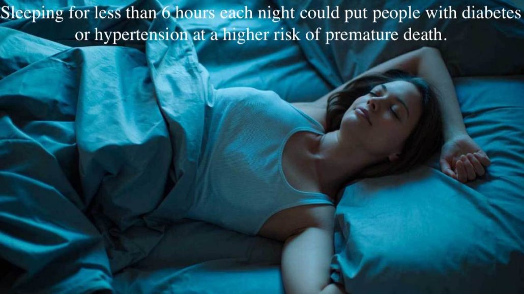
Sleeping for less than 6 hours each night could put people with diabetes or hypertension at a higher risk of premature death.

A new study analyzing the data of more than 1,600 adults found that people with hypertension or type 2 diabetes had a higher risk of death from stroke or heart disease if they slept for less than 6 hours per night.

Type 2 diabetes and hypertension (high blood pressure) are two very common health conditions around the world.

While there are tried and true ways of managing them, these conditions can increase a person’s risk of developing heart disease and experiencing a stroke.

Recently, a study that featured in the Journal of the American Heart Association found that one factor — sleep — may play a significant role for people with these health conditions.

“Our study suggests that achieving normal sleep may be protective for some people with these health conditions and risks,” says lead author Julio Fernandez-Mendoza, Ph.D., from the Pennsylvania State College of Medicine in Hershey.

“However,” he cautions, “further research is needed to examine whether improving and increasing sleep through medical or behavioral therapies can reduce risk of early death.”

Is short sleep duration ‘a useful risk factor?’

In the new study, Fernandez-Mendoza and team analyzed the data of 1,654 participants — 52.5% of whom were women — between the ages of 20 and 74 years. All of the participants had enrolled in the Penn State Adult Cohort.

The researchers split the participants into two categories according to cardiometabolic risk. The participants in one group had stage 2 hypertension or type 2 diabetes, while those in the other group had received a diagnosis of or treatment for heart disease or stroke.

Moreover, the researchers had access to data regarding the participants’ sleep duration, as this cohort had agreed to a 1-night evaluation in a sleep laboratory between 1991 and 1998. They also had access to death records and associated documentation for the years from 1992 through to 2016.

The team’s analysis revealed that of the 512 people who had died by 2016, about two-fifths had died because of causes relating to heart disease or stroke, while close to one-quarter had died following a cancer diagnosis.

What caught the investigators’ attention was the fact that among individuals who had hypertension or type 2 diabetes, the risk of death due to heart disease or stroke was two times higher in those who slept for less than 6 hours per night than in those who slept for 6 hours or more.

For the individuals with one of these two health conditions who slept for longer, the increased risk of premature death was not significant.

Additionally, participants in the heart disease and stroke group who slept for less than 6 hours a night had almost three times the risk of dying from cancer-related causes.

“Short sleep duration should be included as a useful risk factor to predict the long term outcomes of people with these health conditions and as a target of primary and specialized clinical practices.

Julio Fernandez-Mendoza, Ph.D.

“I’d like to see policy changes so that sleep consultations and sleep studies become a more integral part of our healthcare systems. Better identification of people with specific sleep issues would potentially lead to improved prevention, more complete treatment approaches, better long term outcomes, and less healthcare usage,” suggests Fernandez-Mendoza.

While this research adds to the evidence that sleep plays a crucial role in the maintenance of health and well-being, the study authors do admit that their current analysis has some limitations.

The chief limitation, they note, is the fact that they only had access to data on the duration of a single night’s sleep.

As this data came from laboratory observations, they caution that there is a possibility that the participants’ normal sleep patterns may have been different than usual because they were in an unfamiliar environment.

“Nevertheless, the associations found for those other clustered non‐[cardiovascular and cerebrovascular disease] causes of death had the expected [hazard ratios] and provided confidence about the reliability and validity of our findings,” the authors argue in their study paper.

How plant based diets can help people with rheumatoid arthritis

Many recent studies have highlighted the health benefits that a plant based diet can bring. Now, a new review explains just why it can be helpful for people living with rheumatoid arthritis.

Rheumatoid arthritis — a chronic autoimmune condition that causes pain and stiffness in the joints — has a prevalence of between 0.3% and 1% among the world’s population, according to the World Health Organization (WHO).

The condition can become so debilitating as to stop people from continuing in full time work. As the WHO also note, within only 10 years from disease onset, at least 50% of individuals with rheumatoid arthritis in high income countries become “unable to hold down a full time job.”

Doctors usually prescribe a range of drugs and lifestyle adjustments to help people manage their rheumatoid arthritis and make disability less likely. Management strategies that healthcare providers might advise include increased physical activity and weight loss.

Now, a new review appearing in the journal Frontiers in Nutrition shows that following a plant based diet can be a useful intervention when it comes to coping with this condition, as it triggers some helpful biological changes.

‘Symptoms may improve or even disappear’

The review — conducted by specialists from the Physicians Committee for Responsible Medicine in Washington, DC — looked at recent studies that observed the impact of diet on biological mechanisms that are important in rheumatoid arthritis.

It concluded that plant based diets lead to specific changes that can help relieve the symptoms of this condition.

One key way in which plant based diets can be helpful is by reducing levels of inflammation. The review authors cite a study from 2015 that showed participants who ate a plant based diet for 2 months had lower inflammation than those who ate a diet that was high in fat and featured more animal products.

The team also notes that additional research has found an association between adherence to diets high in fat and processed meat and a rise in markers of inflammation. One of these markers is C-reactive protein, a protein present in the blood, and one which reacts to inflammation.

On the other hand, following plant based diets or diets that have a high content of fiber has an association with lower levels of C-reactive protein.

Another study that the review looked at was a randomized clinical trial showing that, after following a low fat vegan diet for 4 weeks, individuals with moderate-to-severe rheumatoid arthritis saw significant improvements in symptoms, including joint pain and stiffness, tenderness, and swelling.

People with rheumatoid arthritis can also benefit from losing extra weight. According to evidence from a 2018 study, overweight individuals with rheumatoid arthritis who shed in excess of 5 kilograms were three times more likely to have improvements in their symptoms compared to those who lost less weight.

The review authors explain that vegetarian and vegan diets appear to help people lose weight, more so than any other diet types.

Finally, the researchers explain that plant based diets also seem to promote a healthy gut environment, since many of these diets are high in fiber, which, as studies have shown, influences the composition of the gut microbiome.

Specifically, plant based diets seem to increase bacterial diversity in the gut, which could help people with rheumatoid arthritis, precisely because they tend to lack bacterial diversity.

The investigators who conducted the review suggest there is a need for further research into the benefits that plant based diets may afford to people with inflammatory autoimmune conditions, as well as their underlying mechanisms.

However, they note that, so far, the emerging evidence suggests that eating more fruits, vegetables, whole grains, and legumes could make a real difference for people with rheumatoid arthritis.

“A plant based diet comprised of fruits, vegetables, grains, and legumes may be tremendously helpful for those with rheumatoid arthritis. This study offers hope that with a simple menu change, joint pain, swelling, and other painful symptoms may improve or even disappear.”

Study co-author Dr. Hana Kahleova

A complex relationship between Alcohol & Dementia

Although alcohol has been popular for millennia, and dementia is increasingly prevalent, scientists are yet to understand the relationship between the two. A recent study sets out for answers.

With dementia predicted to affect 13.9 million adults in the United States by 2060, understanding why these conditions develop is more urgent than ever.

Scientists have uncovered certain factors that increase the risk of developing dementia. Some, such as advancing age, cannot be prevented. However, it is possible to avoid other potential risk factors, such as smoking tobacco.

It is essential to identify modifiable risk factors as understanding these could help prevent or delay the onset of dementia.

Recently, researchers designed a study to look for links between dementia and alcohol consumption in older adults. They published their findings in JAMA Network Open.

What do we already know?

Perhaps surprisingly, as the authors explain, “we know little about the independent associations of quantity and frequency of alcohol consumption with dementia risk.”

Although some studies have investigated alcohol and dementia more broadly, there are still substantial gaps in our understanding. For instance, in an earlier study, researchers calculated alcohol consumption as a daily average across each year.

Using a daily average in this way misses the nuances of alcohol quantity and frequency.

This is an important point. For example, drinking 7 glasses of beer on 1 day each week is likely to have a different impact than drinking 1 glass of beer each night, every day of the week; although the quantity consumed is the same.

The authors of another study concluded that binge drinking in midlife increases the risk of dementia. However, it is still not clear whether regularly drinking smaller amounts of alcohol has the same effect.

Other studies muddy the water further by looking at the links between alcohol, dementia risk, and the presence of apolipoprotein E4 (APOE E4). This gene variant has associations with an increased risk of developing Alzheimer’s.

An earlier study concluded that alcohol consumption increases the risk of dementia and that people with the APOE E4 variant have a higher chance of developing dementia.

Another unknown is how alcohol influences dementia risk for individuals with mild cognitive impairment (MCI). Scientists consider MCI to be the stage between normal age-related cognitive decline and dementia.

Are you looking for alternative health insurance?

Take a short assessment and find affordable short-term health insurance, indemnity insurance, and supplemental health products — like pharmacy benefits and emergency medical plans — to fit your needs.

Alcohol and dementia data

To investigate, the scientists delved into an existing dataset, extracting the information from 3,021 participants, with an average age of 72.

The Ginkgo Evaluation of Memory Study from 2000–2008 supplied the data.

At the beginning of the study, participants provided information about the amount of alcohol they consumed, how often they drank, and what types of drinks they consumed — beer, wine, or liquor.

According to the authors, at the start of the study, each participant went through “a comprehensive neuropsychological battery of 10 tests.” These assessed a range of cognitive functions, and participants completed the tests every 6 months.

Scientists isolated DNA from blood samples to identify carriers of the APOE E4 variant.

Alongside genetic data and details about alcohol use, the scientists also collated information about blood pressure, body weight, height, history of heart disease, and smoking status. They also asked questions to estimate how regularly each participant was involved in social interactions.

Many gaps remain

Of the 3,021 participants, 2,548 did not have MCI, and 473 did have MCI. Over approximately 6 years of follow-up, 512 participants received a diagnosis of dementia.

The authors found that, among those without MCI, no amount of alcohol consumption increased dementia risk compared with those who drank less than one drink per week.

When they analyzed the group of individuals with MCI, it was a similar story; there was no statistically significant difference.

However, dementia risk appeared to be highest for the individuals with MCI who drank 14 drinks per week compared with those who drank less than one drink each week — a relative risk increase of 72%.

The authors also note some differences associated with drinking patterns:

“Daily low-quantity drinking was associated with lower dementia risk than infrequent higher-quantity drinking among participants without MCI at baseline.”

Although the authors are keen to underline the negative impacts of excessive drinking on brain health and health in general, they also note that:

“Our findings provide some reassurance that alcohol consumed within recommended limits was not associated with an increased risk of dementia among older adults with normal baseline cognition.”

When the researchers investigated the potential impact of APOE E4, they found no significant effect, which mirrors several previous studies. The authors theorize that the lack of effect might be because the association is more pronounced in younger populations; they call for further investigation.

In conclusion, the current study provides few solid answers. It confirms, however, that the relationship between alcohol and dementia is complex and likely to require a great deal more research.

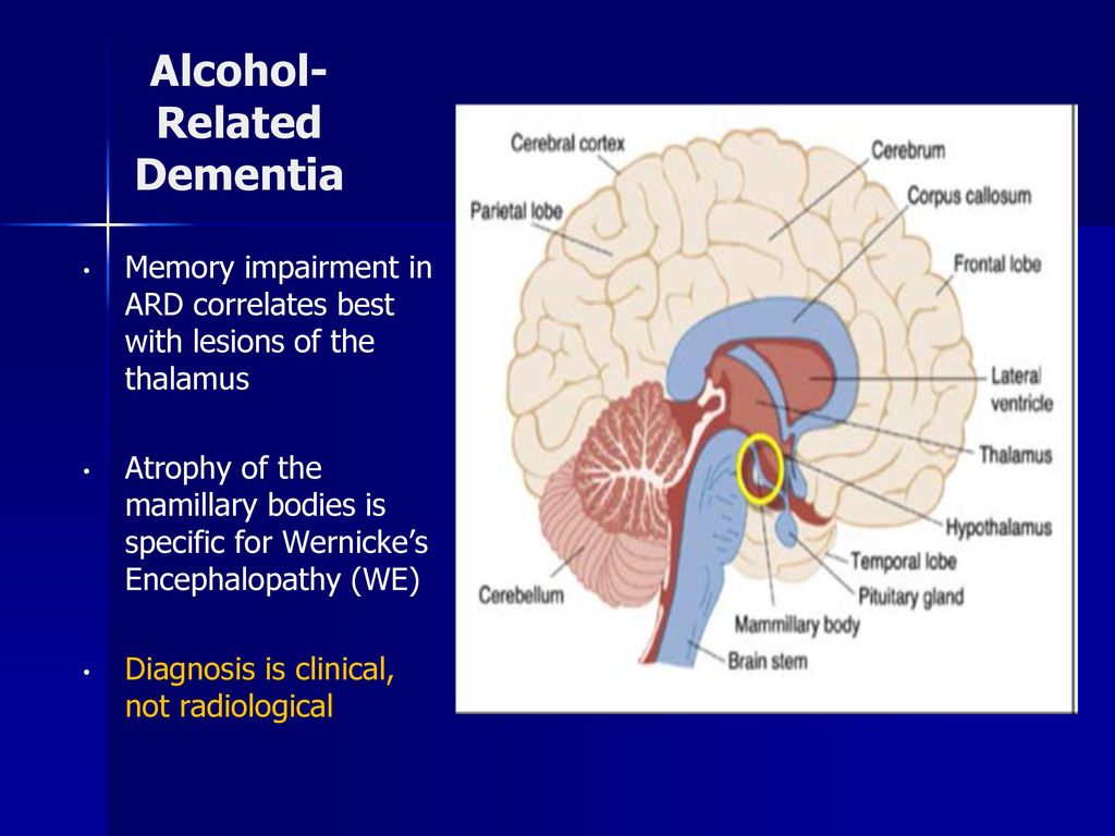
Memory impairment in ARD correlates best with lesions of the thalamus. Atrophy of the mamillary bodies is specific for Wernicke’s Encephalopathy (WE) Diagnosis is clinical, not radiological.