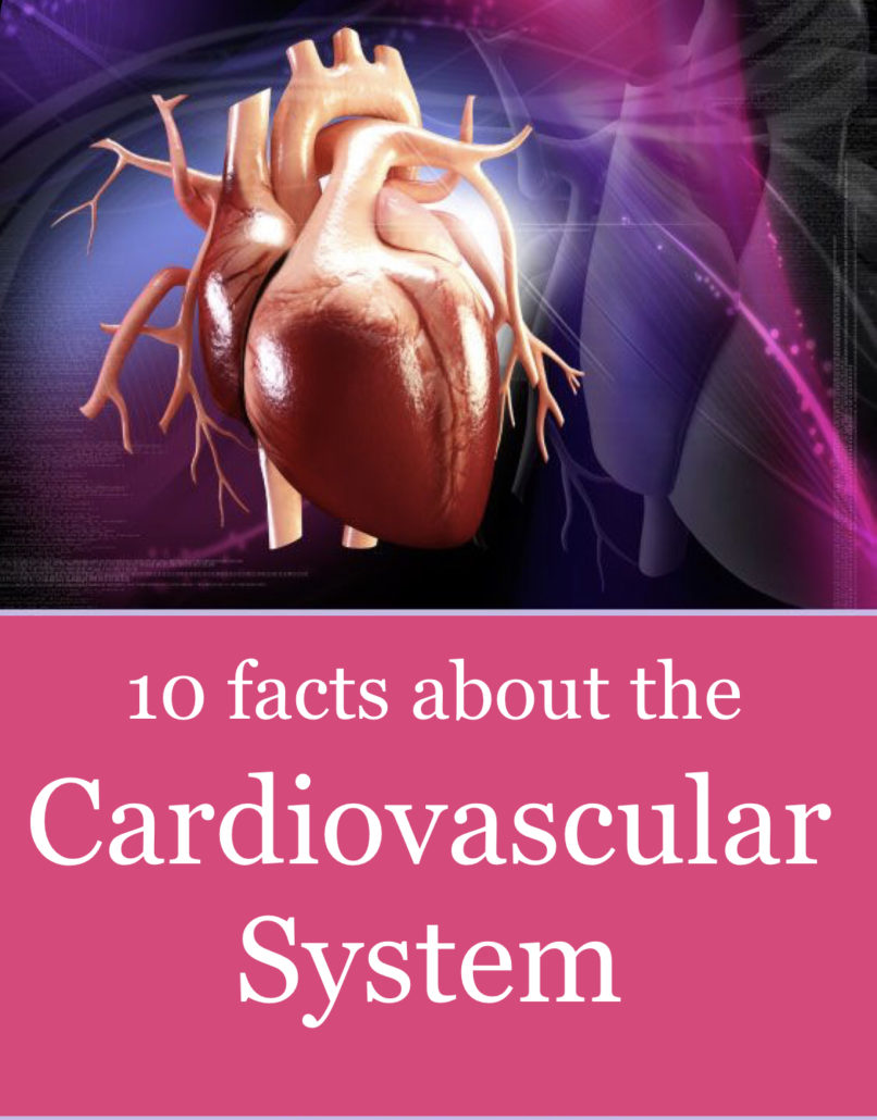
KNOW YOUR HEART. 10 facts about the Cardiovascular system…
The cardiovascular system carries blood and nutrients to the cells of the body. The function of this system has effects on other parts of the body. A nursing student has many facts to learn about the cardiovascular system. Therefore, this article below will start you off with a list of 10 facts about the cardiovascular system that every nursing student should know.
The cardiovascular system or circulatory system consists of the heart, blood and blood vessels. The heart is the pump of the system and sits in the thoracic cavity. The heart sits predominately on the left side. Therefore, approximately two-thirds of the heart is to the left side of the sternum. The blood is a connective tissue. It is the fluid component of the system. The blood is transported to the cells via a network of passageways call the blood vessels.
There are 3 major layers of the heart. The layers are the endocardium, myocardium and the pericardium. The endocardium lines the inner chambers of the heart and the valves. The myocardium makes up the heart wall. The pericardium is the container of the heart. This triple-walled layer protects the heart. Also, the pericardium contains a fibrous layer and a serous layer. The serous layer has two separate layers. These layers are the parietal and visceral layers.
Below is a list of 10 facts about the cardiovascular system that every nursing student should know to help build a foundation of knowledge of the cardiovascular system.
Fact #1: The Cardiovascular System Is A Closed System.
The cardiovascular system is a closed system and also a system which is under pressure. This means if there is a leak in a large vessel it does not drip it sprays, especially a leak on the arterial side.
Therefore, when small leaks occur the system has a method of stopping these leaks. Hemostasis is the system within the blood that stops these leaks. Hemostasis is defined as the stoppage of blood. This system is constantly in action sealing small insults to the system.
Substances contained in the blood assist the process of hemostasis. The blood contains calcium ions and plasma proteins that cause coagulation or clotting within seconds of an injury. These plasma proteins are your clotting factors. (e.g. prothrombin and vitamin K)
Fact #2: The Heart Has Four Chambers.
The heart has four chambers: the right and left atria and the right and left ventricles. The right atrium and right ventricle receive deoxygenated blood from the body. The left atrium and left ventricle receive oxygenated blood from the lungs.
The atria of the heart are mostly reservoirs. The atria only contribute “atrial kick” to the cardiac cycle. Atrial kick or atrial contraction contributes approximately 20% of the volume to ventricular filling.
The ventricles make up the majority of the heart. The ventricles of the heart receive blood from the atrium. They eject blood into the pulmonary system (lungs) and to the systemic circulation (body).
The right side of the heart (ventricle) pumps against a low-pressure system (pulmonary circulation) and the left side of the heart (ventricle) pumps against a high-pressure system (systemic circulation). The left ventricle works harder than all the other chambers because it has to pump against the high pressure of the systemic circulation.
Fact #3: The Heart Has Four Valves.
The right side of the heart contains the tricuspid and pulmonary valves. The left side of the heart contains the bicuspid (mitral) and aortic valves.
The tricuspid and bicuspid (mitral) valves separate the atrium and ventricles. These valves are also called the atrioventricular or AV valves. As you would guess the tricuspid valve had three cusps or leaflets and the bicuspid valve has two cusps or leaflets.
The pulmonary valve opens to the pulmonary circulation and the lungs. The aortic valve opens to the systemic circulation. These valves are also called the semilunar valves because of their shape. The pulmonary and aortic valve each has three cusps or leaflets that are shaped like a half moon.
The “Lub-Dub” sound you hear with your stethoscope is the closing of the heart valves. The heart sound S1 is the closure of the tricuspid and bicuspid (mitral) valves. The heart sound S2 is the closure of the pulmonary and aortic valves.
Fact #4: The Heart Valves Operate Due To A Pressure System.
The valves of the heart open and close due to pressure within the system. During the cardiac cycle, the atria fill with blood. As the atria fill, the pressure in the atria eventually exceeds the pressure in the ventricles. When this happens the tricuspid and bicuspid (mitral) valves open and blood flows into the ventricles.
As blood flows into the ventricles the pressure begins to rise. Eventually, the pressure in the ventricles exceeds the pressure in the atria. This pressure that builds up in the ventricles is attributed to filling volumes of the ventricles. At this time the pulmonary valve and aortic valves close.
Following the isovolumetric contraction of the ventricles, the pulmonary and aortic valves open. Then, blood is ejected into the pulmonary circulation and systemic circulation.
Both the left and right atria fill at the same time and both the left and right ventricles fill at the same time.
Fact #5: Blood Vessels Are The Vascular Portion Of The Cardiovascular System.
Blood vessels include arteries and veins. When the blood leaves the heart it flows into the arteries. Arteries carry oxygenated blood from the heart to systemic circulation. The arteries divide into the smaller arterioles. Next, the arterioles divide into the even smaller capillary network on the arterial side. This arterial capillary network feeds the cells.
The veins divide into smaller venules. The venules divide into the smaller capillary network of the veins. Starting at the capillary network, the capillaries on the vein side pick up carbon dioxide and waste products which travel to the venules and then to the veins and back to the heart.
Blood flows from the heart to the arteries to the arterioles to the arterial capillary network. Then blood moves from the vein capillary network to the venules to the veins and back to the heart.
Fact #6: Blood Vessels Can Constrict And Dilate Having An Effect On Blood Pressure.
The sympathetic nervous system controls the blood vessels. Blood vessels have the ability to constrict or dilate with signals from the sympathetic nervous system.
Vasoconstriction and vasodilation occur when the blood vessels dilate and constrict. Vasoconstriction causes a decrease in the inner diameter of the blood vessel. Vasodilation causes an increase in the inner diameter of the blood vessel.
Blood vessels have an effect on blood pressure.
Remember, blood pressure is the measure of the pressure exerted on the walls of the blood vessel. The greater the pressure within the blood vessel the higher the blood pressure measurement. The lower the pressure within the blood vessel the lower the blood pressure measurement. The systolic pressure is the maximum pressure against the wall of the blood vessel and the diastolic pressure is the recoil.
A change in the diameter of the blood vessels causes changes in the blood pressure. When the blood vessels constrict (vasoconstriction), the blood pressure is higher. This is because the decrease in the diameter of the blood vessel increases the pressure exerted on the lumen. When the blood vessels dilate (vasodilation), the blood pressure is lower. This is because the increase in the diameter of the blood vessel decreases the pressure exerted on the lumen.
Fact #7: The Ventricles Contract Due To Electrical Pathways.
The ventricles contract due to the cardiac conduction system (electrical pathways). The cardiac conduction system consists of the SA or sinoatrial node, the AV or atrioventricular node, the bundle of HIS, the right bundle branch, the left bundle branch and the Purkinje fibers.
The SA node is known as the pacemaker of the heart. It is located on the wall of the right atrium near the entrance to the superior vena cava. The AV node receives electrical impulses from the SA node and transfers them to the bundle of HIS. The bundle of HIS divides into the left and right bundle branch. Impulses travel to each bundle branch down the septum to the Purkinje fibers. The Purkinje fibers innervate the ventricles. The atria of the heart contract before the ventricles.
Fact #8: The Cardiac Cycle Consist of Diastole and Systole.
First of all, the cardiac cycle consists of phases called diastole and systole. These terms should not be confused with the terms diastolic and systolic which refer to blood pressure. These terms are related but not the same. Also, when we talk about diastole and systole we are referring to the ventricles. (e.g. ventricular diastole, ventricular systole)
During systole, when the heart contracts, blood is ejected from the ventricles. The right ventricle ejects blood into the pulmonary circulation (lung) and left ventricle ejects blood into the systemic circulation (body).
During diastole, the heart is at rest and the ventricles are filling. When you think of diastole think of Die, Done, Doing nothing (but filling-ventricular filling) and systole is the opposite.
Fact #9: The Cardiac Cycle Moves Blood Through The Heart.
The phases of the cardiac cycle are diastole and systole. Diastole is divided into early, mid, and late diastole. Systole is divided into early and late systole. Remember, when you think of diastole and systole, think of ventricular diastole and systole. Let’s take a quick walk through diastole and systole. It is easier to begin at diastole.
Early Diastole
Early diastole begins following the closure of the pulmonary and aortic. The tricuspid and bicuspid (mitral) valves are open. During early diastole, the ventricles are rapidly filling. The pressure in the ventricles is beginning to increase.
Mid Diastole
During mid-diastole, the ventricles continue filling but slower. The pressure in the ventricles continues to rise but they still have not exceeded the pressure in the atria. The tricuspid and bicuspid (mitral) valves are still open. The pulmonary and aortic valves close.
Late Diastole
During late diastole, the atrium contract to finish emptying. The atrial contraction is the “atrial kick”. This accounts for approximately 20% of ventricular filling.
Early Systole
At the beginning of early systole, the pressure in the ventricles is greater than the pressure in the atrium. At this time you have an isovolumetric contraction. The ventricular filling and the isovolumetric contraction causes the tricuspid and bicuspid (mitral) to close. This causes the “Lub” sound. The “Lub” is the S1 heart sound.
Late Systole
During late systole, you have ventricular ejection. The blood is ejected into the pulmonary circulation and systemic circulation when the pulmonary and aortic valves are opened. The blood is ejected by the ventricles fast at first then the blood flow slows.
This puts us back to the beginning of early diastole in which the pulmonary and aortic valves close and the tricuspid and mitral valves are open. When the pulmonary and aortic valves close they make the “Dub” sound. The “Dub” is the S2 heart sound. The ventricles are filling during this period.
So, between S1 and S2 you have systole. Between the S2 and the next S1, you have diastole.
Fact #10: There Is A Relationship Between The Cardiac Cycle And Blood Flow.
First of all, the cardiac cycle and blood flow through the heart are very similar. If you understand one you will understand the other. With the cardiac cycle, we move from the top to the bottom (atria to ventricles). With blood flow through the heart, we will move from the right to the left.
Right Atrium
On the venous or return side, deoxygenated blood travel from the venous capillary beds to the venule. Blood continues to travel to the large veins called the superior vena cava and the inferior vena cava. These veins transport blood from the top and bottom of the body. They return blood to the right side of the heart into the right atrium. Then, the right atrium fills causing increased pressure that is eventually greater than the pressure in the right ventricle. This places pressure on the tricuspid valve.
Right Ventricle
The pressure continues to rise until it is greater in the right atrium and causes the tricuspid valve to open. The right ventricle begins to fill. The right atrium contracts causing the final filling of the right ventricle.
As a result of electrical stimulation, the right ventricle contracts. The tricuspid valve closes. The right ventricle ejects blood causing the pulmonary valve to open. Blood enters the pulmonary circulation and moves to the lungs via the pulmonary artery. The blood travels through the capillary bed of the lung. After the blood is oxygenated it returns to the left side of the heart.
Left Atrium
On the left side of the heart, blood travels from the lung to the left atrium via the pulmonary vein. The left atrium begins to fill causing the pressure to increase in the left atrium. The increased pressure is eventually greater in the left atrium producing pressure on the bicuspid (mitral) valve.
Note: If you note above, the pulmonary artery carries deoxygenated blood to the lungs and the pulmonary veins carry oxygenated blood to the left atrium. The pulmonary artery is the only artery in the body that carries deoxygenated blood and the pulmonary vein is the only vein in the body that carries oxygenated blood.
Left Ventricle
The pressure continues to rise until it is greater in the left atrium than the left ventricle and causes the bicuspid (mitral) valve to open. The left ventricle begins to fill. The left atrium contracts causing the final filling of the left ventricle.
Again due to the electrical stimulation, the left ventricle contracts. The bicuspid (mitral) valve closes and the aortic valve opens ejecting blood into the systemic circulation via the aorta. The blood travels throughout the body via the arteries, arterioles to the capillary bed where the process continues.
In conclusion, the list of 10 facts about the cardiovascular system above is by no means all-inclusive. Hopefully, this list will help build a foundation useful in studying the cardiovascular system. Hence, these simple facts will give you a greater understanding of not only how the system works but how it can affect other parts of the body.


