How and when to take a pregnancy test

Pregnancy tests help women to determine if they are pregnant. These tests work by picking up on markers in urine or blood that indicate pregnancy.
Knowing which type of pregnancy test is most suitable, and understanding how to use them, helps ensure results are accurate.
Whether taken at home or in a doctor’s office, being informed about the pregnancy test process makes it easier to understand what the results mean, and what to do next.
Types of pregnancy test
There are two main types of pregnancy test available: urine tests and blood tests.
Urine tests
Urine tests can be taken at home or at the doctor’s office. They detect the presence of the pregnancy hormone, human chorionic gonadotropin (hCG).
The hCG hormone circulates in a woman’s blood once a fertilized egg has attached to the uterus wall. This usually happens 6 days after fertilization.
According to the American Pregnancy Association, levels of hCG double every 2 to 3 days, peak by 8 to 11 weeks of pregnancy, and then begin to level off. HCG can usually be detected by a urine test 12 to 14 days after conception.
Home pregnancy test
Home pregnancy tests can be purchased in a pharmacy or drugstore. They normally cost less than $20 and are easy to use. If used correctly, they give excellent results.
Results are usually visible within 3 to 10 minutes or sometimes sooner.
Clinical urine test
Clinical urine tests are performed at a doctor’s office. Like home tests, they also look for the presence of the hCG hormone in urine.
Although they produce the same level of accuracy, tests performed in a clinical setting usually avoid some of the errors that can happen with home tests.
Blood tests
Although less common than urine tests, blood tests to detect hCG levels are available. These are always carried out at the doctor’s office, and results take longer than urine test results.
Blood tests are usually more expensive but some can be taken earlier than urine tests to determine results. The two types of blood tests are:
* Qualitative hCG blood test: This checks hCG levels, giving a “yes” (pregnant) or “no” (not pregnant) result. They can detect hCG levels around 11 days after conception.
* Quantitative hCG blood test: This is able to measure specific levels of hCG, even if levels are low.
Further tests may be needed if results aren’t as expected. For example, if the hCG level is higher or lower than it should be for the estimated pregnancy stage.
This process can help indicate ectopic pregnancy, miscarriage, or a more accurate due date.
Online tests
Several websites and phone applications have tests that claim to calculate the likelihood of being pregnant. These can be used as a source of information, but should not replace real pregnancy tests.

Who should use a pregnancy test?
Common early indicators of a possible pregnancy include nausea, vomiting, and excessive fatigue.
Any woman of childbearing age, who suspects she may be pregnant, should use a pregnancy test. In particular, women who recently had unprotected sex should consider using a pregnancy test.
Some signs of early pregnancy that should be followed up with a pregnancy test include:
* a missed period
* enlarged, tender, or swollen breasts
* fatigue
* increased urination
* nausea
* vomiting

How to use pregnancy tests
For a clinical urine test, the doctor will ask for a sample of urine. This can be tested immediately.
Blood tests require a sample of blood to be drawn and sent for testing.
Home pregnancy tests are simple to use. It is important to follow the instructions closely to get the most accurate result.
Most brands can be used on the first day of a missed period, but some more sensitive tests can be used sooner. Some clinical tests can also be taken earlier than this.
In general, home pregnancy urine tests involve one of the following methods:
* urinating directly onto the test stick
* urinating into a cup and dipping the test stick in
* urinating into a cup and using a dropper to drop urine onto the test window
Many manufacturers recommend taking two home tests to confirm results. This is because hCG levels may be too low to be detected at first. To ensure the most accurate results, it is best to wait a few days to 1 week between the two tests.

Accuracy
Many home pregnancy tests claim to be accurate up to 99 percent of the time. Clinical tests can be even more accurate.
However, using them correctly is vital. Several studies suggest incorrect use of tests has led to many women reporting false results.
Most tests can be used on the first day of a missed period but it’s advisable to wait 1 week after a missed period for accuracy.
This is because 10 to 20 percent of pregnant women won’t detect their pregnancy on the first day of their missed period. This can be due to many women having irregular periods, or miscalculating when their period is due.
In addition, a number of other factors play a role in the accuracy of results, including:
* time of day the test is taken
* length of time after conception
* when ovulation occurs
* how quickly the fertilized egg implants
* how well instructions are followed
* how sensitive the test is

What do the results mean?
If a home pregnancy test reads positive, it is important for a doctor to confirm the results with a clinical test.
Results of a pregnancy test are either positive or negative. If taking a clinical test, the doctor will explain what the results mean. If using a home test, it’s important that women refer to the test instructions to ensure they understand what the colors, symbols, or words indicate.
Positive result
A positive result indicates pregnancy, even if the color, symbol, or words on the test window are faint. It’s a good idea to confirm this result by taking a clinical test.
Very rarely, a false-positive result occurs. This means the woman taking the test is not pregnant, despite the test indicating she is. False-positives can occur due to:
* early miscarriage
* certain medications
* ovarian tumors
* liver disease
* kidney failure
* expired test kits
Negative result
A negative result means pregnancy is unlikely. However, false-negative results are common.
These are where pregnancy may still occur but is not yet showing up on the test. They can occur for a number of reasons, including:
* incorrectly taking the test
* testing too soon
* testing with urine that is too diluted
* not waiting long enough for results
* expired or faulty test
Retesting within a week is advisable for all test results. This is particularly important if a woman is experiencing some of the symptoms of early pregnancy or if the test result is negative.
When to see a doctor
A woman should always consult her doctor if she has any queries about the pregnancy or test results. If the home pregnancy test is positive, a doctor can confirm this with a clinical test and begin to arrange prenatal care.
If a home pregnancy test is negative but a woman’s periods don’t resume, she should consult a doctor to determine the cause of the missed periods.

Early-life asthma may contribute to childhood obesity

Asthma affects millions of children in the United States, and so does obesity. A new study may have found a link between the two, as early-life asthma may contribute to the development of childhood obesity.

New research suggests that asthma may increase the risk of childhood obesity.
It is estimated that asthma affects around 1 in 10 U.S. children.
As for childhood obesity, the Centers for Disease Control and Prevention (CDC) report that 12.7 million children in the U.S. – or approximately 17 percent – are obese.
Childhood obesity and asthma are often found to occur together, but existing research has not yet clarified whether asthma actually contributes to the childhood obesity “epidemic.”
New research from the University of Southern California (USC) in Los Angeles aims to fill this research gap by examining the effects of asthma and asthma medication on childhood obesity.

Assessing the link between early-life asthma and childhood obesity
The researchers examined the medical records of 2,171 non-obese children between 5-8 years old.
These children were enrolled in the Southern California Children’s Health Study (CHS) and were clinically followed for 10 years.
CHS is a large-scale, long-term, detailed study of the lasting effects of air pollution on the respiratory and metabolic health of children.
The USC researchers examined 10 years’ worth of data collected in the CHS, and they also performed a replication analysis on an independent sample of 2,684 CHS children. This latter cohort was followed from the average age of 9.7 years to 17.8 years.
During this time, researchers measured the children’s height and weight annually, categorizing them into normal, overweight, and obese. Children were deemed overweight or obese if their body mass index (BMI) was at or above the 85th and 95th percentile, respectively, compared with the BMI standards set by the CDC.
Researchers team assessed the children’s asthma using the physician’s diagnosis as reported by the children or by their parents.
The parents also filled in complex questionnaires on sociodemographic factors and smoking exposure at home or in utero, as well as physical activity patterns and history of respiratory illness.
The scientists used Cox regression to evaluate the associations between asthma and the incidence of obesity during the follow-up period.
Children with asthma 51 percent more likely to become obese
At the beginning of the study, over 18 percent of the children were overweight and over 13 percent had been diagnosed with asthma.
During the follow-up period, 15.8 percent of children developed obesity.
After statistical adjustments, the researchers found that early childhood asthma did contribute to the development of obesity in subsequent years, mainly during early childhood and adolescence.
Compared with non-obese, asthma-free children, the non-obese children who did have asthma were 51 percent more likely to develop obesity during follow-up. The results stayed the same following sociodemographic adjustments and adjustments for other variables.
Additionally, children with a history of wheezing were at a 42 percent higher risk of developing obesity.
The age of asthma onset did not seem to influence the obesity risk.
Regarding the link between the use of asthma medications and obesity, the researchers made another interesting discovery. They found that the use of asthma rescue medications, such as an asthma inhaler, significantly lowered the risk of obesity. These results were independent of physical activity.

Significance of the study
The authors note that theirs is one of the few studies to link early-life asthma with an increased risk of developing obesity.
Some of the study’s limitations include the fact that the information was self-reported, and that the scientists did not have access to sufficient information regarding diet or physical activity patterns.
Frank Gilliland, the study’s senior author, points out that a combination of asthma and obesity can trigger other metabolic diseases in adulthood, including prediabetes and type 2 diabetes.
Children who have asthma are often overweight or obese, but the scientific literature has not been able to say asthma causes obesity.
However, our study and that of others support the finding that having asthma in early childhood may lead to increased risk of childhood obesity.

Although it could be that the respiratory problems prevent children with asthma from playing and exercising as much as healthy children, it is interesting, the authors note, that this study accounted for physical activity yet still showed these results.
Early diagnosis and treatment of asthma may help prevent the childhood obesity epidemic. Part of the problem may be a vicious cycle where asthma and obesity negatively affect each other. Our results also suggest that asthma inhalers may help prevent obesity in children. Although this observation warrants further study, it is interesting that the correlation exists irrespective of physical activity and other asthma medication use.

Arthritis and physical activity

Just 45 minutes of exercise a week can benefit older adults with arthritis

Engaging in physical activity can reduce pain and help to maintain mobility and independence for older adults with arthritis, but current exercise recommendations are often unachievable for this population. Now, however, a new study finds that exercising for just 45 minutes is enough to reap the benefits.

Researchers say that 45 minutes of moderate-intensity exercise per week can benefit older adults with arthritis.

#Arthritis is a term used to describe inflammation of the joints. Osteoarthritis (OA) is the most common form of arthritis, caused by “wear and tear” of cartilage that protects the joints. As the cartilage breaks down, pain, swelling, and joint movement problems may occur.
Older adults are most commonly affected by arthritis. According to the Centers for Disease Control and Prevention (CDC), between 2010-2012, approximately 49.7 percent of adults aged 65 and older in the United States reported having been diagnosed with arthritis by a doctor.
Arthritis and other rheumatic conditions are a leading cause of disability among U.S. adults; around 9.8 percent of those with doctor-diagnosed arthritis in 2010-2012 reported limitations in activity as a result of the condition.
Arthritis and physical activity
While physical activity can be challenging for many older adults with arthritis, it can help patients better manage their condition and maintain physical functioning. In addition, exercise can reduce the risk of other health problems, such as type 2 diabetes and heart disease.
The Physical Activity Guidelines for Americans recommend that adults aged 65 and older engage in 150 minutes of moderate-intensity aerobic activity each week.
However, according to our last study only 1 in 10 older U.S. adults with knee OA meet these recommendations. This is most likely because they find such guidelines unachievable with their condition.
With this in mind, the team set out to determine whether older arthritis patients might be able to benefit from lower levels of physical activity than the current recommendations.
To reach their findings, the researchers analyzed the data of 1,629 adults aged 49 and older who were part of the Osteoarthritis Initiative – a nationwide study that aims to identify prevention and treatment strategies for patients with knee OA. All participants had pain, aching, or stiffness in joints of the hips, knees, or feet.
The physical functioning of each subject was assessed at study baseline and 2 years later through self-reported outcomes. Physical activity and functioning were also measured using movement-monitoring accelerometers.
‘Even a little activity is better than none’
After 2 years, the physical functioning of around a third of participants had either improved or remained high, the team reports.
It was the participants who engaged in regular exercise that experienced higher physical function. However, subjects did not need to meet the recommendation of 150 minutes of moderate-intensity activity each week to see benefits.
The team found that older adults who engaged in just 45 minutes of moderate-intensity activity, such as brisk walking, per week were 80 percent more likely to maintain or improve their physical functioning than those who exercised for under 45 minutes weekly.
The researchers say that their findings applied to both men and women with arthritis of the lower joints.
We found the most effective type of activity to maintain or improve your function 2 years later was moderate activity, and it did not need to be done in sessions lasting 10 minutes or more, as recommended by federal guidelines.
Participants who engaged in more than 45 minutes of moderate-intensity activity every week saw greater benefits to physical function, but the researchers believe that their findings show that older adults with arthritis need only perform a third of the recommended activity to remain functional.
Even a little activity is better than none. For those older people suffering from arthritis who are minimally active, a 45-minute minimum might feel more realistic.

How reducing alcohol intake can benefit health

As 2017 arrived, many of us are thinking about the healthy lifestyle changes we want to make for the coming year. Do you plan to exercise more? Lose weight? Quit smoking? How about cutting down on alcohol intake? While that last question may have prompted a highly resistant shake of the head from many readers who enjoy their after-work beer or a glass of wine with dinner, there is no better time to review your alcohol intake.
Are you planning to reduce your alcohol intake for 2017? If so, it could yield numerous health benefits.
During the month of January, millions of people will be giving up alcohol as part of a campaign dubbed “Dry January.”
Popularized by British organization Alcohol Concern, Dry January aims to “change the conversation” about alcohol by encouraging people to stop drinking for 1 month.
You might think that quitting the drink for just 31 days is unlikely to have a big impact on health, but many participants who take part in Dry January report better sleep, increased energy, and weight loss.
Most importantly, abstaining from alcohol for 1 month may encourage a reduction in alcohol intake in the longer term. A study published in the journal Health Psychology in March found that people who took part in Dry January showed a decrease in alcohol consumption over the subsequent 6 months.
In this article, we take a closer look at how alcohol affects the body, as well as the health benefits of reducing alcohol intake.
The scale of drinking in the U.S.
The Dietary Guidelines for Americans 2015-2020 recommend that adults who drink alcohol should only do so in moderation – that is, up to one alcoholic beverage a day for women and two for men.
However, the National Institute on Alcohol Abuse and Alcoholism (NIAAA) report that in 2014, almost a quarter of adults in the United States engaged in binge drinking – consumption of around four drinks for women and five for men in a 2-hour period – over the past month.
Additionally, a further 6.7 percent reported engaging in heavy drinking over the past month, which is defined as consuming at least five drinks on one occasion for 5 or more days over the past 30 days.
Over the holidays and other special occasions, it can be hard to say no to that glass of bubbly when everyone else is celebrating with a drink in hand. Some of us may even see alcohol as a reward for a hard week at work or as a stress-relieving aid.
That bottle of beer or glass of wine might feel good in the moment, but if consumed in high amounts or too often, it could wreak havoc on your physical and mental well-being.
The effects of alcohol intake

The brain
Those of you who drink are likely to have experienced the dreaded hangover at least once. Feelings of sickness, dizziness, dehydration, and headache are all caused by drinking too much the night before.
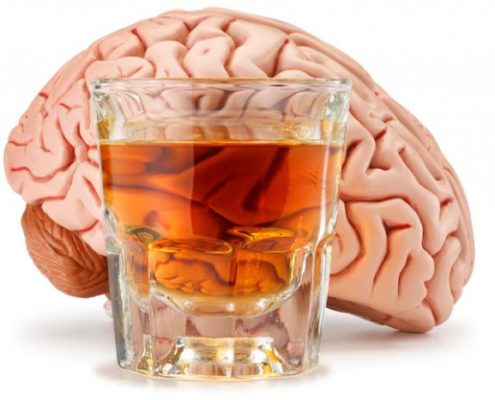
Alcohol can cause an imbalance of neurotransmitters in the brain.
However, symptoms of excess alcohol intake can begin long before a hangover. In fact, alcohol can cause problems soon after the first sip.
The NIAAA explain that alcohol disrupts communication between nerve cells in the brain by causing an imbalance in the levels of neurotransmitters – the chemicals that relay signals from one nerve cell to another.
An imbalance of neurotransmitters can cause changes to mood, behavior, and coordination that are characteristic of excess alcohol intake.
Researchers have found that the brain can adapt to changes in neurotransmitter levels caused by alcohol, but this is not necessarily good news. An adjustment such as this enables us to build tolerance to alcohol, and it may fuel the development of alcohol use disorders.
In 2014, around 16.3 million adults in the U.S. had an alcohol use disorder, and in 2010, alcohol abuse cost the country around $249 billion.
The heart
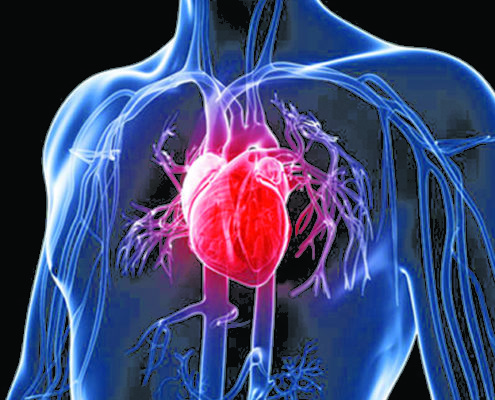
According to the American Heart Association, excess alcohol consumption can increase levels of fats in the blood, which are called triglycerides. High triglyceride levels are known to contribute to the buildup of plaque in the arteries, known as atherosclerosis, which can raise the risk of heart attack and stroke.
Drinking too much – particularly over a long period of time – can also lead to high blood pressure, arrhythmia (irregular heartbeat), cardiomyopathy (enlargement of the heart muscle), and stroke.
It should be mentioned, however, that an increasing number of studies have suggested that moderate drinking may actually benefit heart health. A study reported by Medical News, linked moderate alcohol intake to reduced risk of heart attack and heart failure.
Still, some researchers have questioned the heart health benefits of moderate drinking, and health organizations recommend not initiating alcohol consumption solely to reap such – as yet unproven – rewards.
The liver
When we drink alcohol, the liver breaks it down so that it can be eliminated from the body. Drinking too much over time, however, can cause the liver to become seriously damaged.
In 2011, almost half of liver cirrhosis deaths in the U.S. were related to alcohol.
Heavy alcohol intake can lead to alcoholic fatty liver disease – characterized by a buildup of fat in the arteries – alcoholic hepatitis (inflammation of the liver), and alcoholic cirrhosis (severe liver scarring and structural damage).
The latter is the most advanced form of liver injury caused by heavy alcohol use; according to the NIAAA, of all cirrhosis deaths in the U.S. in 2011, around 48 percent were related to alcohol intake.
The pancreas
The pancreas is crucial for digestion and energy production; it sends enzymes to the small intestine to break down carbohydrates, proteins, and fats.
However, drinking alcohol excessively over a long period of time can disrupt pancreatic function; instead of enzymes being sent to the small intestine to aid digestion, they are secreted into the pancreas itself.
This can lead to pancreatitis, which is characterized by inflammation and swelling of the blood vessels in the pancreas. Around 5 percent of people with alcohol dependence develop the condition.
Alcohol and cancer
An increasing number of studies have associated even light to moderate alcohol intake with increased risk of certain cancers.
A recent study reported by MNT found that each glass of white wine consumed daily may raise the risk of melanoma by 13 percent, while an earlier study linked low alcohol intake to a greater risk of breast cancer.
Other cancers that have been associated with alcohol use include mouth, esophageal, throat, and liver cancers.
The health benefits of reducing alcohol intake
Of course, many of the health risks associated with alcohol intake are related to excessive, long-term drinking. However, even those who drink lightly and in moderation could benefit from reducing their alcohol intake.
Weight loss
Alcoholic beverages are full of calories; a standard glass of white wine contains around 121 calories, while a pint of beer contains at least 150 calories. Needless to say, just a few drinks per week can contribute to weight gain.
Reducing alcohol intake may help you to lose weight.
However, this can be turned around by reducing alcohol consumption; a survey from Alcohol Concern found that around 49 percent of people who take part in Dry January reported weight loss, as well as increased energy.
The NIAAA’s alcohol calorie calculator can help you work out how many calories you consume from alcoholic beverages each week.
Better mood
A drink or two on a Saturday night may put you in good spirits in the short term, but the effect is unlikely to last.
Alcohol intake – especially in large quantities – can interfere with the neurotransmitters that regulate mood, increasing the risk of anxiety, stress, and depression.
As such, cutting back on the liquor means that you are more likely to be in a happier state of mind.
A better night’s sleep
Studies have shown that alcohol can disrupt sleep. Research reported by MNT in 2014, for example, found that drinking can interfere with the body’s ability to regulate sleepiness and wakefulness, which can lead to insomnia.
Dr. Shawn Adhami recommends steering clear of drinking just before bedtime, as this can disrupt normal sleep patterns.
Deep sleep is when the body restores itself, and alcohol can interfere with this,” he explains. As the alcohol starts to wear off, your body can come out of deep sleep and back into REM [rapid eye movement] sleep, which is much easier to wake from. That’s why you often wake up after just a few hours sleep when you’ve been drinking.
It is no surprise, then, that cutting down on drinking can mean a better night’s sleep; a survey from Alcohol Concern found that 62 percent of participants who took part in Dry January reported sleep improvements.
A healthier stomach
Even a little alcohol can irritate the stomach, increasing its production of acid.
This can lead to inflammation of the stomach lining, known as gastritis, causing symptoms such as stomach pain, vomiting, and diarrhea. Heavy drinking may even cause stomach bleeding.
By reducing alcohol intake, such problems can be reduced or avoided.
Ready to cut down on drinking?
If cutting back on your alcohol intake has now made it on to your list of New Year’s resolutions, these tips from the NIAAA can help you get started:
* Monitor how much you are drinking: the NIAAA recommend carrying a drinking tracker card, allowing you to track every alcoholic beverage consumed
* Set a goal: do you want to give up alcohol completely, or just limit alcohol intake to once or twice a week? Set yourself a goal of how much you want to drink and when. The NIAAA recommend having some alcohol-free days
* Avoid drinking “triggers”: if you are more likely to drink around certain people or environments, try to avoid such scenarios
* Learn to say “no”: it can be hard to turn down a drink when offered one, especially on special occasions, but the NIAAA recommend having a polite “no, thank you” at the ready. The organization’s module on building drink refusal skills may help
* Pace yourself: aim to consume no more than one standard alcoholic beverage per hour when drinking, sip it slowly, and make every other drink a nonalcoholic one such as water or juice
* Avoid drinking on an empty stomach: eating food alongside an alcoholic beverage means that the alcohol will be absorbed into the bloodstream more slowly.

Dr Afshin Shawn Adhami

Stroke vs. Aneurysm: Treatment Options

A brain aneurysm is a bulge in an artery in the brain that has the potential to burst or rupture. A ruptured aneurysm can cause a type of stroke called a subarachnoid hemorrhage.
An estimated 5 percent of Americans may have or develop a brain aneurysm each year, according to the Mayfield Clinic.
Not all aneurysms cause stroke, and vice-versa. However, if a person is at risk for a burst aneurysm, treatment is often required to prevent this potentially life-threatening occurrence.
Doctors don’t detect an estimated 85 percent of aneurysms until they burst. Fortunately, if a person can seek them quickly enough, there are treatments available for both stroke and aneurysm.
There are different forms of stroke and aneurysm. The treatment that a doctor gives will depend on what type occurs.

Types of stroke
A stroke may occur due to either the blood supply to the brain being blocked or a blood vessel in the brain rupturing.
Two stroke types exist: hemorrhagic and ischemic.
Hemorrhagic strokes account for an estimated 13 percent of all stroke types, according to the American Heart Association. This stroke type occurs when a vessel in the brain bursts.
As a result, blood spills into the brain. The added blood can compress the brain, which can keep life-saving oxygen and blood from getting to the brain tissue.
Hemorrhagic strokes are usually the result of one of two causes: an aneurysm or a collection of abnormal blood vessels in the brain that can rupture.
Ischemic strokes are those that result from a blockage in an artery in the brain. When a blood clot breaks free from its place in an artery, it can lodge in a portion of the brain. This keeps blood from flowing freely to the brain.
Without the oxygen and nutrients that the blood brings to the brain tissue, the tissue dies. The result can be impaired body functioning or death.

Types of brain aneurysm
An aneurysm is a weakened area of an artery in the brain. This area of weakness can result in bulging as blood flows through it. If enough pressure is generated against this weak area, it can burst and blood will flow out.
Doctors may classify an aneurysm by the symptoms it causes. These include:

Aneurysm that hasn’t burst
An aneurysm that hasn’t burst can range in size from large to small. Some people may have a small brain aneurysm and go their entire lives without knowing it.
If an aneurysm gets bigger, it can start to press on areas around the brain. This can cause symptoms such as:
* Pain behind the eye
* Dilated pupil
* Drooping eyelid
* Numbness or weakness on one side of the face
* Pain behind the eye
* Visual changes, such as seeing double
Some people have a family history of brain aneurysms, and they may undergo imaging studies to help a doctor identify if the person may have one that hasn’t burst. Most will largely go undiagnosed until a person starts experiencing symptoms or the aneurysm bursts.
Doctors may classify aneurysms that haven’t burst by their location or size in the brain. These include:
* Fusiform: An aneurysm that has no distinct “neck” and bulges in every direction
* Giant: An aneurysm that is larger than 2.5 centimeters in diameter
* Saccular: Also known as a “berry” aneurysm, this aneurysm type has a distinct neck and typically bulges to one side of an artery

‘Leaking’ aneurysm
A leaking aneurysm is one that may have enough of a weak area to allow a small amount of blood through. This leak can lead to a sudden, severe headache that indicates the extra pressure on the brain.
A leaking aneurysm is dangerous because it is often a precursor to a burst aneurysm.

Burst aneurysm
A burst aneurysm may require immediate brain surgery.
A burst aneurysm occurs when the weakened area of an aneurysm ruptures, leaking a significant amount of blood.
The symptoms associated with a burst aneurysm are severe, and the side effects can be deadly. They include:
* Confusion
* Nausea and vomiting
* Neck stiffness
* Passing out
* Sudden, severe headache
* Visual changes, such as blurred or double vision
A burst aneurysm is a type of subarachnoid hemorrhage. This is because the bleeding occurs between the brain and the membrane that covers the brain. A subarachnoid hemorrhage is a cause of a hemorrhagic stroke.
According to Johns Hopkins Medicine, an estimated 3 percent of all strokes are subarachnoid hemorrhage strokes.
Treatment for brain aneurysm
If a person has a burst aneurysm and seeks emergency medical attention, medications are often given to prevent symptoms and damage from getting worse.
Examples of immediate medical treatments include:
* Anti-seizure medications to prevent seizures that can occur
* Pain medications to relieve the very intense headache a ruptured aneurysm can cause
* Vasodilators, which are medicines that cause blood vessels to expand
Vasodilators keep the affected vessel from tightening too much and stopping important blood flow. These medications also keep a person’s blood pressure from getting too high, which could lead to increased pressure in the skull and bleeding.
Doctors will take into account many factors when determining a potential surgical approach to aneurysm treatment.
Doctors don’t always treat an aneurysm that hasn’t burst, but usually will when it is causing symptoms. Burst aneurysms require fast treatment. Examples of considerations are the patient’s symptoms, health history, and the aneurysm’s size and location.
Several treatment approaches exist for brain aneurysms.

Surgical clipping
This involves accessing the area in the brain where the aneurysm occurred, placing a metal clip at the “neck” of the aneurysm. This keeps blood from flowing into and out of the aneurysm.
The surgical clipping approach involves removing a piece of the skull to access this portion of the brain.

Endovascular coiling
Endovascular coiling is a less invasive approach to aneurysm treatment. It involves accessing a large artery in the body, usually the femoral artery in the groin. A small tube is threaded through this artery up to the area where the aneurysm is.
A doctor will then introduce a special kind of metal coil or glue that fills the aneurysm and blocks blood flow. Clipping and coiling are both known as endovascular treatments.

Flow diversion with stents
This approach involves inserting a special device to re-route blood flow past the aneurysm. The flow diversion procedure can only be performed on an aneurysm that hasn’t burst.
The ability to do this reduces one of the greatest risks associated with surgery, which is accessing the aneurysm itself. With time, the aneurysm should close because it doesn’t receive blood flow that causes it to bulge.

Artery occlusion or bypass

Medical attention should be sought if any symptoms of a brain aneurysm are suspected.
An approach to use if an aneurysm can’t be coiled or clipped. This involves a surgeon removing a portion of the skull to access the area of the aneurysm. An artery from the patient or a donor artery is sewn to the damaged artery, re-routing blood.
If a person experiences a burst aneurysm and subarachnoid hemorrhage, they are at increased risk for an occurrence known as a vasospasm.
A vasospasm is when an artery in the brain tightens or narrows. This limits blood flow and causes symptoms like confusion, restlessness, and muscle weakness.

Treatment for stroke
Treatments for stroke depend upon the stroke type. Doctors can treat an ischemic stroke with a medication called tissue plasminogen activator (tPA). If given within several hours of the first symptoms, this medicine can help to break up a clot and improve blood flow.
If a person cannot receive tPA, another option is retrieving a clot through a procedure known as a mechanical thrombectomy. This involves using a special device to remove the clot and insert a stent to keep the artery open.
A doctor will usually access the clot in a similar method to endovascular coiling, which is through an artery in the groin.
Hemorrhagic stroke treatments due to a subarachnoid hemorrhage are the same as those listed above for an aneurysm. Treatments for a hemorrhagic stroke caused by a collection of abnormal blood vessels may also involve clipping or bypass surgery to prevent bleeding.
According to University Hospital in Newark, NJ, the fatality rates for hemorrhagic stroke are higher than ischemic stroke. The effects of the massive bleeding associated with a hemorrhagic stroke are often difficult to combat, even with today’s technological advancements.
As a result, it’s important that people who have any symptoms of an aneurysm seek immediate medical treatment.

senior man with headache

senior man with headache

Eating fiber keeps gut microbes from eating you

The gut microbiota plays a crucial role in maintaining our body’s overall health. New research shows what happens if we do not feed our gut microbes with the fiber they need to survive.

Gut bacteria are essential to keeping our body healthy.

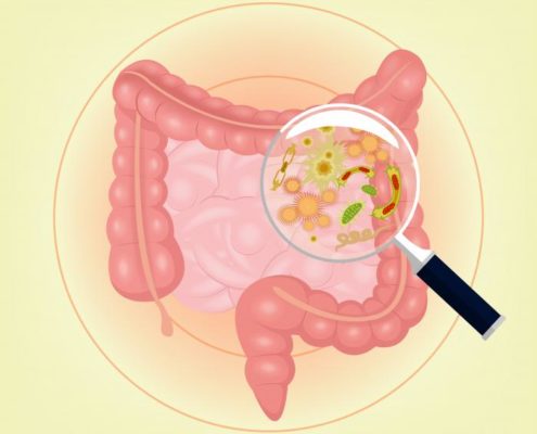
Our gut microbiota contains at least 1,000 different species of known bacteria, summing up 3 million genes.
We share one third of our gut bacteria with other people, while the composition in our other two thirds is unique to each one of us.
Gut microbiota is important to our health because it contributes to a healthy immune system by acting as a barrier against other harmful microorganisms
It also helps with digesting foods that the stomach and small intestine have not been able to digest, as well as producing some vitamins.

We have always been told by healthcare professionals and nutritionists that fiber is important to a healthy diet.
But new research examines exactly what happens if our intestinal microbes do not receive the appropriate amount of fiber.

Studying the behavior of gut bacteria in mice

Researchers bred mice especially for the study. Mice were born and raised without any gut bacteria of their own. They then received a transplant of 14 bacteria that normally live in the human gut.
Knowing the genetic signature of each bacterium, scientists were able to track the evolution of each one of them over time. They used a germ-free lab facility and genetic techniques that allowed them to see what bacteria were present and active under different dietary conditions.
Researchers infected the mice with a strain of bacteria that is the equivalent of E. coli in humans. Then they examined the impact of diets with varying amounts of fiber, as well as a diet with no fiber at all.
Researchers tried a diet that was 15 percent fiber, made from minimally-processed grains and plants. They also tried a diet that was rich in prebiotic fiber – a purified form of soluble fiber that is similar to what some processed foods and dietary supplements contain.

Gut microbes really need their fiber

As revealed by the study – published in the journal Cell – the induced infection did not fully spread in mice that received the 15 percent-fiber diet. Their mucus layer remained thick, protecting them against the infection.
But when scientists replaced the diet with one that lacked fiber altogether, gut microbes started eating the mucus. Even a few days of fiber deprivation led the bacteria to start invading the colon wall.
Gut microbes rely on fiber for their food, and when they do not get it, they start eating away at your gut. This makes the gut more prone to infections.
The diet rich in supplement-like prebiotic fiber had the same results as the diet lacking fiber completely. The mucus layer started eroding as a result of the action of microbes.

Researchers were also able to see what fiber-digesting enzymes the bacteria were making. They found 1,600 different enzymes that digest carbohydrates – a complexity similar to the one found in the human gut.
A lack of fiber also triggered a higher production of such mucus-degrading enzymes.
Scientists were able to look at images of the “goblet” cells on the colon wall that produce mucus. They could clearly see how the mucus layer got progressively thinner as the mice received less fiber.
In a normal gut, mucus is being produced and degraded at a steady pace. But on a fiber-deprived diet, mucus was degraded at a much higher pace than it was produced.
Examining the gut tissue of infected mice, researchers were able to see inflammation across a wide area of thinning, and even patchy tissue.
Infected mice that received a diet rich in fiber also displayed inflammation but across a much smaller area.

Future research to study different diets In the future, we want to study the effect of different prebiotic combinations over a longer period of time, as well as the impact of an intermittent natural fiber diet.
Researchers would also like to find the biomarkers that signal the state of the mucus layer in human guts, such as the number of mucus-degrading bacteria.
We would like to study the impact of a low-fiber diet on chronic illnesses such as inflammatory bowel disease.
“While this work was in mice, the take-home message for humans amplifies everything that doctors and nutritionists have been telling us for decades: Eat a lot of fiber from diverse natural sources.
Your diet directly influences your microbiota, and from there it may influence the status of your gut’s mucus layer and tendency toward disease. But it’s an open question of whether we can cure our cultural lack of fiber with something more purified and easy to ingest than a lot of broccoli.”intestinal-bacteria-under-a-magnifying-glass